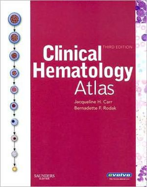
Clinical Hematology Atlas
Ideal for identifying cells at the microscope, this atlas covers the basics of hematologic morphology, including examination of the peripheral blood smear, basic maturation of the blood cell lines, and discussions of a variety of clinical disorders. Over 400 photographs, schematic diagrams, and electron micrographs illustrate hematology from normal cell maturation to the development of various pathologies.\ • Numerous illustrations include excellent schematic diagrams, photomicrographs, and...
Search in google:
Ideal for identifying cells at the microscope, this atlas covers the basics of hematologic morphology, including examination of the peripheral blood smear, basic maturation of the blood cell lines, and discussions of a variety of clinical disorders. Over 400 photographs, schematic diagrams, and electron micrographs illustrate hematology from normal cell maturation to the development of various pathologies.• Numerous illustrations include excellent schematic diagrams, photomicrographs, and electron micrographs. • Introductory chapters succinctly describe the peripheral blood smear, its preparation and examination, and hematopoiesis in general. • Coverage of cellular maturation includes schematics that illustrate the maturation of each cell line individually and highlight the cell in question. • Descriptions for each cell type include size, reference intervals, and nuclear and cytoplasmic characteristics. • Body Fluids chapter covers the other fluids found in the body besides blood, using images from cytocentrifuged specimens. • White blood cell differential table shows cells found in a normal white blood cell differential. • Overview of hematopoiesis includes a schematic drawing along with a detailed presentation of each cell, demonstrating the relationship between individual stages of hematopoiesis and the overall development scheme. • Morphologic abnormalities are presented in chapters on erythrocytes and leukocytes, along with a schematic description of each cell, to provide correlations to various disease states. • Coverage of common cytochemical stains, along with a summary chart for interpretation, aids in classifying malignant and benign leukoproliferative disorders. • Spiral binding and a compact size make this book easy to use in a laboratory setting.• NEW Normal Newborn Peripheral Blood Morphology chapter covers the normal cells found in neonatal blood. • More examples of specific cells and disorders allow you to compare abnormal cells to each other and to normal cells, differentiating those that are similar. • Expanded Evolve resources include case studies, study questions, links to related websites, and content updates. Michele D. Raible This small atlas covers the basics of hematologic morphology in 23 chapters, including examination of the peripheral blood smear, basic maturation of the blood cells lines, and discussions of a variety of clinical disorders. The atlas is offered as a source to be used when cells have to be identified (i.e., at the microscope), and is intended to be used in conjunction with a textbook of hematology. The authors state that they designed the volume for a diverse audience that includes clinical laboratory science students, medical students, residents, and practitioners. The introductory chapters succinctly describe the peripheral blood smear, its preparation and examination, and hematopoiesis in general. The chapters on cellular maturation include schematics that illustrate the maturation of each cell line individually and highlight the cell in question. A color print and electron micrograph are included, along with a description of each cell that includes size, reference intervals, and nuclear and cytoplasmic characteristics. Additional chapters contain information on morphology in a variety of disease states and clinical conditions. Illustrations and photomicrographs are generally quite good and the descriptions are well organized and concise. Tables are included to briefly summarize information throughout. This small volume emphasizes morphology for practical identification of cells while at the microscope. The authors intend it to be used with a textbook that addresses physiology and diagnosis, along with morphology. Several aspects of the book are useful, including the emphasis of the relationship of individual cells to hematopoiesis, concise descriptions, electron micrographcorrelations, and generally fine quality photomicrographs. This atlas appears best suited to beginning clinical laboratory medicine students, medical students, or clinical laboratory practitioners retraining in hematology; the book lacks the detail that more experienced hematology professionals may desire and is generally available in more comprehensive hematology atlases.
Ch. 1Introduction to peripheral blood smear examination1Ch. 2Hematopoiesis11Ch. 3Erythroid maturation19Ch. 4Megakaryocyte maturation33Ch. 5Myeloid maturation41Ch. 6Monocyte maturation55Ch. 7Eosinophil maturation65Ch. 8Basophil maturation75Ch. 9Lymphoid maturation81Ch. 10Variations in size and hemoglobin content of erythrocytes91Ch. 11Variations in shape and color of erythrocytes95Ch. 12Inclusions in erythrocytes109Ch. 13Diseases affecting erythrocytes117Ch. 14Nuclear alterations of leukocytes133Ch. 15Cytoplasmic alterations of leukocytes137Ch. 16Acute myeloid leukemia151Ch. 17Acute lymphoid leukemia165Ch. 18Myeloproliferative disorders169Ch. 19Myelodysplastic syndromes175Ch. 20Malignant lymphoprolifertive disorders185Ch. 21Cytochemical stains197Ch. 22Microorganisms205Ch. 23Miscellaneous cells213Ch. 24Body fluids223
\ From The CriticsReviewer: Valerie L. Ng, PhD MD(Alameda County Medical Center/Highland Hospital)\ Description: This is the third edition of a color atlas of cells and other items found in peripheral blood and body fluids. New to this edition is the spiral-binding — invaluable for keeping the book open when referring to a picture. The previous edition was published in 2004.\ Purpose: The purpose is to provide a handy bench guide as a quick and readily accessible reference for morphological identification. This book has nicely met the authors' objective.\ Audience: This book is intended to have a wide appeal to anyone interested in blood or body fluid morphology: medical students, residents and fellows in any medical specialty, practicing clinicians, allied health providers, etc. The main beneficiaries will be clinical laboratory scientists, pathologists and clinical hematologists — students or practitioners. \ Features: My first impression of this book was, "Wow, this is beautiful." It is primarily pictures, the clarity and color reproduction of which are superb. The book is divided into sections starting with normal hematopoiesis, normal maturation, leukemias, myelodysplasias, lymphoma, microorganisms, miscellaneous cells, newborn blood and body fluids. All pictures depict Wright-stained preparations. Each picture has a short descriptive section highlighting key morphological features and associated disease states. The descriptions are purposely short, as this atlas is intended to be a companion to a textbook (Rodak et al, Hematology: Clinical Principles and Applications, 3rd edition (Elsevier, 2007)) in which physiology and diagnosis are addressed. The inclusion of various body fluids (synovial, pleural, CSF) make this a hybrid of atlases covering only peripheral blood and those covering only body fluids. There are deficiencies in this hybrid approach, however. For example, there is no depiction of cells in CSFs of neonates — a morphologically difficult specimen for those not used to seeing neonatal CSF. Similarly, other fluids commonly evaluated in the hematology lab (i.e., semen, pericardial, ascites, urine) are not included. Regardless, the entities displayed in this atlas cover about 90 percent of what the average bench clinical laboratory scientist will see in the course of a career.\ Assessment: This is a beautiful atlas that will be used every day by the bench clinical laboratory scientist. Get it.\ \ \ \ \ Michele D. RaibleThis small atlas covers the basics of hematologic morphology in 23 chapters, including examination of the peripheral blood smear, basic maturation of the blood cells lines, and discussions of a variety of clinical disorders. The atlas is offered as a source to be used when cells have to be identified (i.e., at the microscope), and is intended to be used in conjunction with a textbook of hematology. The authors state that they designed the volume for a diverse audience that includes clinical laboratory science students, medical students, residents, and practitioners. The introductory chapters succinctly describe the peripheral blood smear, its preparation and examination, and hematopoiesis in general. The chapters on cellular maturation include schematics that illustrate the maturation of each cell line individually and highlight the cell in question. A color print and electron micrograph are included, along with a description of each cell that includes size, reference intervals, and nuclear and cytoplasmic characteristics. Additional chapters contain information on morphology in a variety of disease states and clinical conditions. Illustrations and photomicrographs are generally quite good and the descriptions are well organized and concise. Tables are included to briefly summarize information throughout. This small volume emphasizes morphology for practical identification of cells while at the microscope. The authors intend it to be used with a textbook that addresses physiology and diagnosis, along with morphology. Several aspects of the book are useful, including the emphasis of the relationship of individual cells to hematopoiesis, concise descriptions, electron micrographcorrelations, and generally fine quality photomicrographs. This atlas appears best suited to beginning clinical laboratory medicine students, medical students, or clinical laboratory practitioners retraining in hematology; the book lacks the detail that more experienced hematology professionals may desire and is generally available in more comprehensive hematology atlases.\ \ \ 3 Stars from Doody\ \
